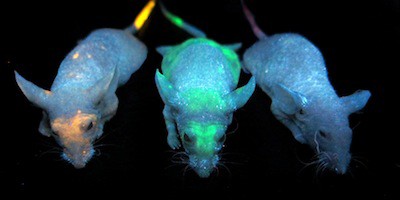
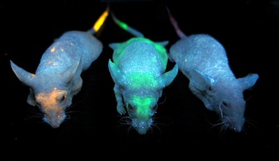
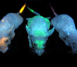
Mice injected with quantum dots – nanocrystals make of cadmium sulfide, zinc and other materials – glow under UV light. A new study shows that the concentration of nanoparticles in skin is a good proxy for how much has accumulated in other organs, like the liver or the spleen. (Credit: Edward A. Sykes & Qin Dai)
Researchers have come up with a quick, non-invasive way to measure nanoparticle exposure: examine the skin. Previous work with gold nanoparticles and quantum dots made of various metals has shown that these nanoparticles accumulate primarily in the liver or the spleen, but they show up in the skin too; in high enough doses, they can even change the skin’s colour.
The latest study shows that levels in skin are directly correlated with those in the liver and spleen, so a skin sample could provide a non-invasive of measuring an animal’s exposure to nanoparticles. The method could also work for nanosilver, an emerging environmental contaminant.
Original research paper published in the the journal Nature Communications on May 13, 2014.
Names and affiliations of selected authors