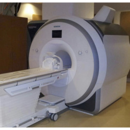
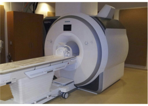
Researchers from the University of Western Ontario developed a new tool using this 3T Siemens MRI system to track multiple sclerosis progression in patients. (Photo Credit: Ravi Menon)
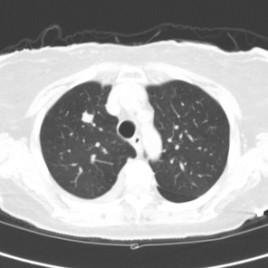
A new tool provides a means to track multiple sclerosis (MS) progression and could lead to early diagnosis, a new study shows. The method – called Quantitative Susceptibility Magnetic Resonance Imaging (MRI) – allows researchers to track small changes in the magnetic field of the brain caused by iron distribution and white matter lesions that are linked to MS.
They found that lesions appeared in the same areas in all MS patients which tells researchers where to look for the earliest possible diagnosis.
This technique could be readily extended to other hospitals with MRI scanners, but it could take at least 5 years for it to be used as a valid MS diagnostic tool.
Original research paper published in the the journal Radiology on May 4, 2014.
Names and affiliations of selected authors